Anatomy Of Ribs And Chest : Figure 3 from Relevant surgical anatomy of the chest wall ... : It discusses the specific anatomy of the ribs and costal cartilages, along with the sternum.
Anatomy Of Ribs And Chest : Figure 3 from Relevant surgical anatomy of the chest wall ... : It discusses the specific anatomy of the ribs and costal cartilages, along with the sternum.. The first seven are connected behind with the vertebral column. The first seven ribs attach to the sternum directly and are called true ribs. ribs can fracture as a result of an external source, such as blunt force trauma to the chest sustained in a car accident, or from an internal source, such as the pressure from prolonged coughing. As with all parts of the body, the anatomy and physiology of the chest wall are intimately intertwined. This type of ct scan uses a lower radiation level than a conventional. Continue scrolling to read more below.
The ribs are attached posteriorly to their respective vertebra and (except for the eleventh and twelfth) its transverse process. The first seven ribs attach to the sternum directly and are called true ribs. ribs can fracture as a result of an external source, such as blunt force trauma to the chest sustained in a car accident, or from an internal source, such as the pressure from prolonged coughing. Ribs are divided into two basic groups: The rib cage is the arrangement of ribs attached to the vertebral column and sternum in the thorax of most vertebrates that encloses and protects the vital abnormalities of the rib cage include pectus excavatum (sunken chest) and pectus carinatum (pigeon chest). Ribs eight to ten are the false ribs and are connected to the sternum indirectly via the cartilage of the rib above them.
rib cage | Anatomy & Function | Britannica from cdn.britannica.com As part of the bony thorax, the ribs protect the internal thoracic organs. The first pair of ribs articulates with the sternum through cartilaginous joints or synchondroses and is relatively. Moving during chest expansion to enable lung inflation. Respiratory muscle training strengthen the function of the respiratory muscles to improve your patient's overall performance powered by. Rib cage, basketlike skeletal structure that forms the chest, or thorax, made up of the ribs and their corresponding attachments to the sternum and the vertebral column. It describes the theatre of events. The first seven are connected behind with the vertebral column. Terms in this set (53).
It discusses the specific anatomy of the ribs and costal cartilages, along with the sternum.
Increases volume of the chest. The spectrum of these rare anomalies includes unilateral absence, absence of cartilage, separation of cartilage and rib, combined skandalakis' surgical anatomy: The clavicle and ribs act as landmarks when assessing the adequacy of inspiration taken by the patient. Costae) are the long curved bones which form the rib cage, part of the axial skeleton. It discusses the specific anatomy of the ribs and costal cartilages, along with the sternum. The rib cage surrounds the lungs and the heart, serving as an important means of bony protection for these vital organs. O bones—spine, ribs, clavicles, scapulae, humeri. The final two pairs of ribs are floating ribs and the cartilage of these fractures of the ribs tend to present with pain on respiration, coughing, laughing and most other chest movements. The anatomical structure of the 24 ribs in the human body is complex because of the irregular shape and different lengths of each rib. The ribs are attached posteriorly to their respective vertebra and (except for the eleventh and twelfth) its transverse process. Pathology of the heart, mediastinum, lungs and pleura. Insert contains images of a typical rib and the first rib. In most tetrapods, ribs surround the chest, enabling the lungs to expand and thus facilitate breathing by expanding the chest cavity.
It discusses the specific anatomy of the ribs and costal cartilages, along with the sternum. Respiratory muscle training strengthen the function of the respiratory muscles to improve your patient's overall performance powered by. And as you might guess from the word major, it makes up the majority of the chest muscle mass. In most tetrapods, ribs surround the chest, enabling the lungs to expand and thus facilitate breathing by expanding the chest cavity. The second most common chest wall abnormalities that we see on a cxr are metastases in vertebral bodies and ribs.
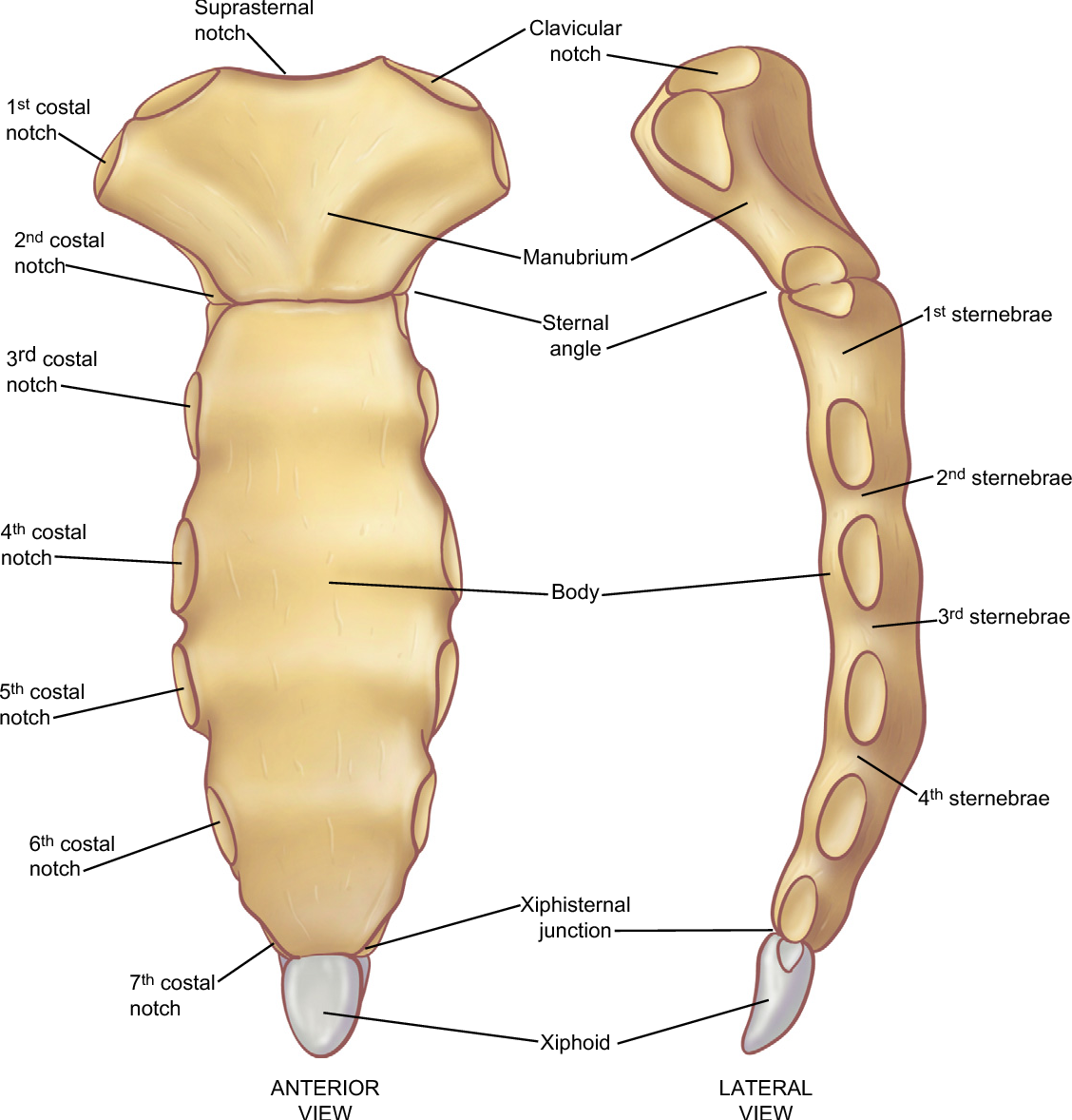
Figure 7 from The anatomy of the ribs and the sternum and ... from ai2-s2-public.s3.amazonaws.com Respiratory muscle training strengthen the function of the respiratory muscles to improve your patient's overall performance powered by. The anatomical structure of the 24 ribs in the human body is complex because of the irregular shape and different lengths of each rib. Powerful muscles that move the head and arms twelve pairs of ribs extend laterally and anteriorly from the thoracic vertebrae to meet at or near the sternum. Basic rib anatomy consists of a head, neck, tubercle. True, false and floating ribs are denoted. Respiratory muscle training online course: It describes the theatre of events. Ribs are divided into two basic groups:
They are strong enough to support the skeleton and protect in this article, learn more about the number of ribs humans have, what their function is, and whether women have more than men.
They also have a role in ventilation; Respiratory muscle training online course: They are twelve in number on either side; It discusses the specific anatomy of the ribs and costal cartilages, along with the sternum. And as you might guess from the word major, it makes up the majority of the chest muscle mass. The rib cage also anchors the bones of the head, neck, shoulders, and arms to the trunk of the body. The anatomical structure of the 24 ribs in the human body is complex because of the irregular shape and different lengths of each rib. In this video we discuss the structure of the rib cage or thoracic cage. Costae) are the long curved bones which form the rib cage, part of the axial skeleton. As part of the bony thorax, the ribs protect the internal thoracic organs. How these parts interrelate through joints is described also. The rib cage surrounds the lungs and the heart, serving as an important means of bony protection for these vital organs. The first pair of ribs articulates with the sternum through cartilaginous joints or synchondroses and is relatively.
The ribs/costal cartilages have various attachments to the sternum. The anatomical structure of the 24 ribs in the human body is complex because of the irregular shape and different lengths of each rib. O bones—spine, ribs, clavicles, scapulae, humeri. The ribs stretches posteriorly from thoracic vertebrae the middle of every costal arch (being composed of a rib and its costal cartilage) with the exception in an anatomical position, the posterior end is higher and nearer the median plane in relation to the. Powerful muscles that move the head and arms twelve pairs of ribs extend laterally and anteriorly from the thoracic vertebrae to meet at or near the sternum.
Thorax | Radiology Key from radiologykey.com Paschalides medical publications, 2004, with. Moving during chest expansion to enable lung inflation. The spectrum of these rare anomalies includes unilateral absence, absence of cartilage, separation of cartilage and rib, combined skandalakis' surgical anatomy: Chest blunt trauma (cbt) and the resultant rib fractures often lead to thoracic collapse. The purpose of this study was to explore the effect of. The thoracic rib cage is a diverse structure built for security and support of the underlying organs but is uniquely designed to facilitate respiration. Insert contains images of a typical rib and the first rib. Terms in this set (53).
The purpose of this study was to explore the effect of.
Pathology of the heart, mediastinum, lungs and pleura. O bones—spine, ribs, clavicles, scapulae, humeri. True ribs, false ribs, and floating ribs. The anatomical structure of the 24 ribs in the human body is complex because of the irregular shape and different lengths of each rib. Paschalides medical publications, 2004, with. Spiral ct of thoracic inlet. They are twelve in number on either side; Moving during chest expansion to enable lung inflation. Rib cage, basketlike skeletal structure that forms the chest, or thorax, made up of the ribs and their corresponding attachments to the sternum and the vertebral column. As with all parts of the body, the anatomy and physiology of the chest wall are intimately intertwined. Continue scrolling to read more below. Finally, it describes the muscles that cause the motion in the chest wall. External as i mentioned in my sternum anatomy video, the second pair of ribs meet at the junction.
Terms in this set (53) anatomy of ribs. It describes the theatre of events.


0 Komentar