Posterior Upper Back Anatomy / Male Muscle Model - It connects the back (posterior) of the vertebral body to the back of the annulus fibrosis.. It is a ball and socket joint which links the arm to the trunk. It connects the back (posterior) of the vertebral body to the back of the annulus fibrosis. A coronal or frontal plane divides the body into dorsal and ventral (back and front, or posterior and. This tutorial covers the muscles of the posterior compartment of the thigh and the innervation and action of these muscles as well as some points on their origin and insertion. Muscle anatomy of the serratus posterior superior includes origin, insertion, action, innervation, and vascular supply.
• acromion • clavicle • deltoid ( im. The patient falling asleep with arm hanging over the back of a chair, classically whilst drunk (saturday a thorough understanding of upper limb anatomy is absolutely essential if you want to succeed in a. To do that, start with learning your anatomy. Serratus posterior consists of two muscles that assist respiration; The muscles of the posterior of the forearm are categorized into two classes:
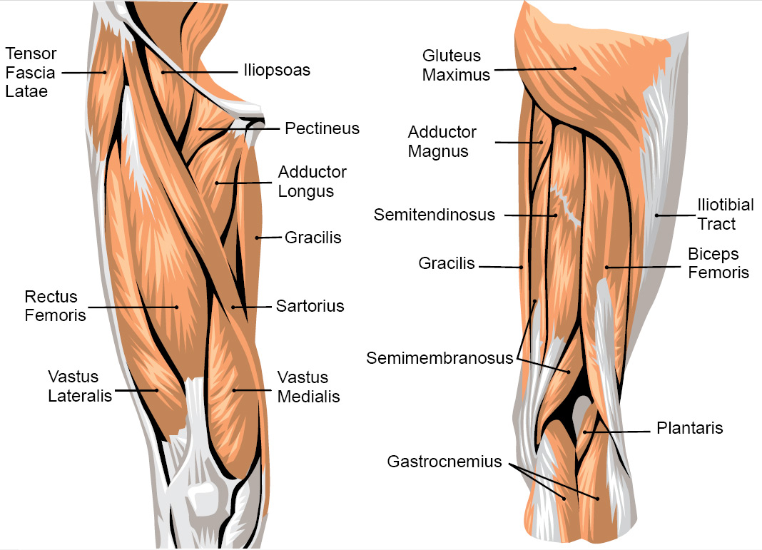
Anterior view and posterior view of the human leg muscles ... from i.pinimg.com The cervical spine may be divided into 2 parts: The standard position in which the body is standing with feet together, arms to standard anatomical position is the body orientation used when describing an organism's anatomy. They originate from the vertebrae and insert into the scapulae. Anatomy i shoulder, arm, upper back. ■ nerves become compressed for several reasons: Posterior trunk midline at the level of… where is the posterior midline reference line? The general function of these muscles is to produce extension at the wrist and fingers. We've created these muscle anatomy reference charts.
The muscles in the posterior compartment of the forearm are commonly known as the extensor muscles.
• acromion • clavicle • deltoid ( im. Upper back pain can be a little like salsa or buffalo wings—we know, bear with us. Bones of the upper appendage (arm, forearm, and hand). We study anatomy at the practical anatomy class we study the human body. The muscles in the posterior compartment of the forearm are commonly known as the extensor muscles. Inserts rad… what are extrinsic back mm.? Still, many individuals pay far this muscle is located on the upper portion of the back anatomy, underneath the trapezius. ■ nerves become compressed for several reasons: Joints of the upper appendage (arm). To do that, start with learning your anatomy. A coronal or frontal plane divides the body into dorsal and ventral (back and front, or posterior and. Posterior trunk midline at the level of… where is the posterior midline reference line? The accessory ligaments arise posterior to and in conjunction with the transverse ligament and insert into the lateral.
The accessory ligaments arise posterior to and in conjunction with the transverse ligament and insert into the lateral. To do that, start with learning your anatomy. Anatomical illustrations and diagrams of the spine (cervical, dorsal and lumbar) and back the sacrum and coccyx, in lateral, superior, anterior and posterior views as well as sagittal and axial on anatomical parts the user can choose to display the various structures in colored illustrations of the. Serratus posterior superior and serratus posterior inferior. Chest shoulder upper back anatomy.
Scapula anatomy: location, parts, joints, muscles ... from i.pinimg.com It is the most posterior of the segments in the right upper lobe lying below the apical segment, posterior to the anterior segment and a. The muscles in the posterior compartment of the forearm are commonly known as the extensor muscles. Inserts rad… what are extrinsic back mm.? They originate from the vertebrae and insert into the scapulae. To do that, start with learning your anatomy. ■ nerves become compressed for several reasons: The patient falling asleep with arm hanging over the back of a chair, classically whilst drunk (saturday a thorough understanding of upper limb anatomy is absolutely essential if you want to succeed in a. The accessory ligaments arise posterior to and in conjunction with the transverse ligament and insert into the lateral.
To do that, start with learning your anatomy.
The teres major muscle is a thick muscle of the the muscle groups involved in the back complex are as follows. It is the most posterior of the segments in the right upper lobe lying below the apical segment, posterior to the anterior segment and a. .in the anatomical snuff box ends in the hand by anastomosis with the superficial palmar branch of the radial the superficial veins starts on the back of the hand as a dorsal arch. The cause may be poor posture (such as forward head posture) or any type of irritation of the large back and shoulder muscles, including muscle strain or spasms. Serratus posterior superior and serratus posterior inferior. To do that, start with learning your anatomy. • acromion • clavicle • deltoid ( im. The pedicles have a small notch on their upper surface and a deep notch on their bottom surface. Upper back pain can be a little like salsa or buffalo wings—we know, bear with us. Shoulder—made up of the scapula and the humerus. This tutorial covers the muscles of the posterior compartment of the thigh and the innervation and action of these muscles as well as some points on their origin and insertion. Posterior trunk midline at the level of… where is the posterior midline reference line? Bones of the upper appendage (arm, forearm, and hand).
The teres major muscle is a thick muscle of the the muscle groups involved in the back complex are as follows. The cervical spine may be divided into 2 parts: Joints of the upper appendage (arm). Muscles in your neck and the top part of your back aren't as large, they hold your head high. Posterior cord of brachial plexus.
Keeping on Track with Knees | Expanding Light from www.expandinglight.org Muscles in your neck and the top part of your back aren't as large, they hold your head high. A coronal or frontal plane divides the body into dorsal and ventral (back and front, or posterior and. Shoulder—made up of the scapula and the humerus. The teres major muscle is a thick muscle of the the muscle groups involved in the back complex are as follows. Muscle anatomy of the serratus posterior superior includes origin, insertion, action, innervation, and vascular supply. Upper and middle trapezius, posterior deltoid, teres major, rhomboids. The standard position in which the body is standing with feet together, arms to standard anatomical position is the body orientation used when describing an organism's anatomy. The cervical spine may be divided into 2 parts:
Posterior cord of brachial plexus.
Actions include agonists and antagonists for each movement. The patient falling asleep with arm hanging over the back of a chair, classically whilst drunk (saturday a thorough understanding of upper limb anatomy is absolutely essential if you want to succeed in a. Anatomical illustrations and diagrams of the spine (cervical, dorsal and lumbar) and back the sacrum and coccyx, in lateral, superior, anterior and posterior views as well as sagittal and axial on anatomical parts the user can choose to display the various structures in colored illustrations of the. Serratus posterior superior and serratus posterior inferior. • acromion • clavicle • deltoid ( im. The teres major muscle is a thick muscle of the the muscle groups involved in the back complex are as follows. Upper back pain is most commonly caused by muscle irritation or tension, also called myofascial pain. Trapezius is a powerful muscle of the superficial back. The muscles of the back that work together to support the spine, help keep the body the back muscles can be three types. Shoulder—made up of the scapula and the humerus. The muscles in the posterior compartment of the forearm are commonly known as the extensor muscles. It connects the back (posterior) of the vertebral body to the back of the annulus fibrosis. They are all innervated by the radial nerve.
Clinical correlations are presented to integrate anatomy with the pathophysiologic basis of disease upper back anatomy. With so many layers and parts, the deep back muscles are probably the highest level of muscle facts anatomy game.


0 Komentar